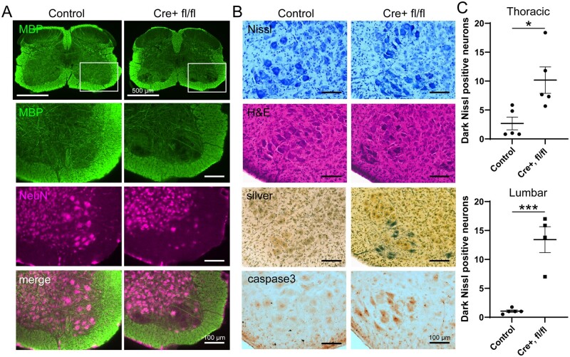
Figure 5.
Degeneration of ventral horn motor neurons in the spinal cord of VPS35 cKO mice crossed with Synapsin-1-Cre mice at P12. (A) Immunofluorescence co-localization of myelin basic protein (MBP) and NeuN in the lumbar spinal cord of VPS35fl/fl/Cre or control (VPS35fl/fl or VPS35fl/wt) mice. Representative high magnification images are shown from the boxed region of the ventral horn (upper panel). The loss of large NeuN-positive motor neurons and associated MBP-positive fibre tracts in VPS35fl/fl/Cre mice is indicated. (B) Representative histological staining of lumbar spinal cord (ventral horn) sections with cresyl violet (Nissl) and haematoxylin/eosin, in addition to Gallyas silver stain or immunohistochemical labelling of activated caspase-3, from VPS35fl/fl/Cre or control (VPS35fl/fl or VPS35fl/wt) mice. Notice that large ventral horn motor neurons display condensed dark staining for Nissl and haematoxylin/eosin with abnormal morphology, and stain positive for silver and activated caspase-3 in VPS35fl/fl/Cre mice. (C) Quantitative counts of dark, condensed Nissl-positive neurons in thoracic and lumbar spinal cord sections from VPS35fl/fl/Cre mice (n = 5 mice) and control mice (VPS35fl/fl, VPS35fl/wt, VPS35wtlwt; n = 5 mice) sampled across 5–6 sections per animal. Bars indicate the mean ± SEM. *P < 0.05 or ***P < 0.001 by unpaired, two-tailed Student’s t-test. Scale bars: 100 or 500 μm, as indicated.

