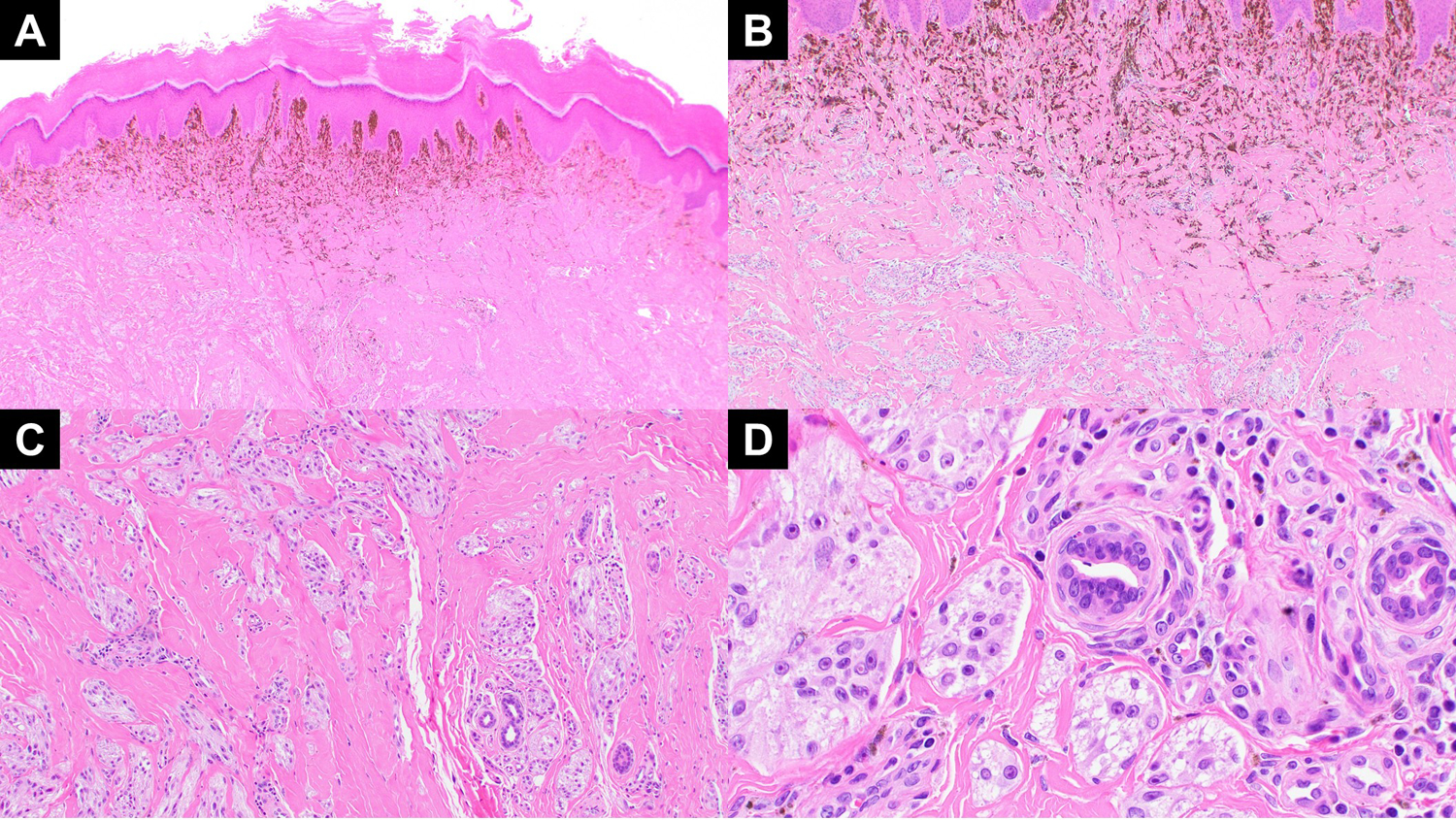
Figure 2:

(A) A low power view of case 2 showing nodular melanocytic lesion with prominent aggregation of melanophages in the subepithelial region. (B) Highlights the top heavy melanization and plexiform arrangement of the dermal melanocytes in a sclerotic stroma. (C) The dermal melanocytes also show aggregation around the neurovascular bundles in the dermis. (D) Higher magnification view of variably dermal nests of melanocytes with moderate atypia and Spitzoid and epithelioid cytomorphology.
