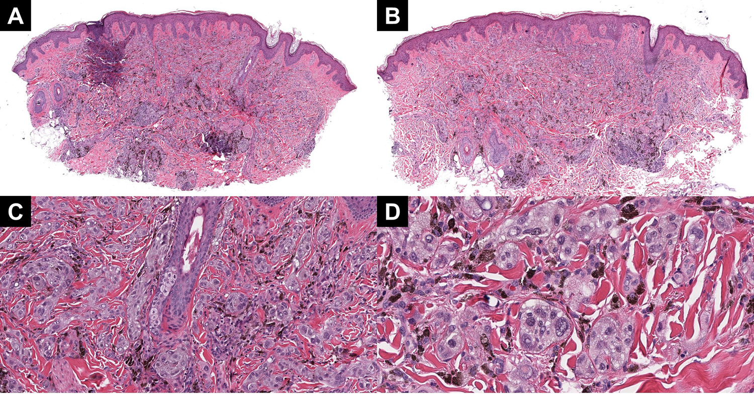
Figure 3:

(A and B) Two profiles of case 3, highlighting the plexiform growth pattern and extensive pigmentation throughout the lesion. (C) On higher power, clustering around the adnexa and neurovascular bundle is notable. (D) The cytology is epithelioid and definitively Spitzoid, with multiply vacuolated nuclei and high-grade atypia.
