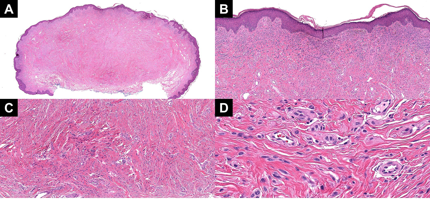
Figure 4:

(A) A low power image showing a melanocytic tumor in a highly sclerotic stroma. (B) Superficially, the tumor showed a more nested profile. (C) With descent, the tumor developed into smaller nests and single units of Spitzoid melanocytes in a sclerotic stroma. (D) A high power view of the dispersion to single units and small nests of Spitzoid melanocytes near the base.
