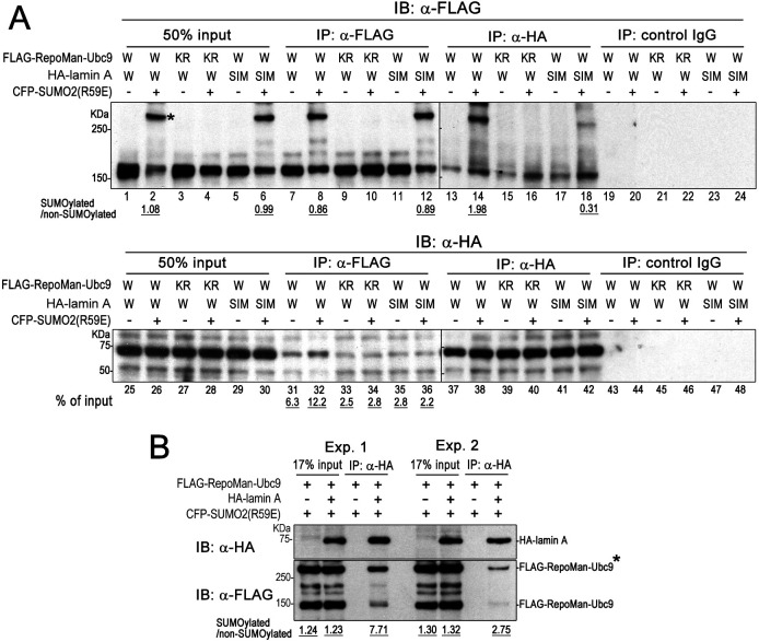
Fig. 5.
SUMOylation of RepoMan enhances the interaction between RepoMan and lamin A. (A) 293FT cells were transfected with plasmids expressing HA–lamin A(WT) (W) or HA–lamin A[SIM3(AA)] (SIM), FLAG–RepoMan(WT)–Ubc9 (W) or FLAG-RepoMan(K762R)-Ubc9 (KR), and CFP–SUMO-2(R59E) as indicated and subjected to immunoprecipitation (IP) using an anti-FLAG antibody, anti-HA antibody or normal mouse IgG. FLAG–RepoMan with or without SUMOylation and HA–lamin A in input and immunoprecipitated samples were detected using anti-FLAG and anti-HA antibodies, respectively. The asterisk indicates the CFP–SUMO-2 modified FLAG–RepoMan–Ubc9. Relative amounts of SUMOylated RepoMan and non-SUMOylated RepoMan in each sample were determined by quantifying the signals of immunoblots (IB) using ImageJ. The amounts of HA–lamin A precipitated using anti-FLAG antibody were quantified from signals of triplicate experiments. (B) Quantification of RepoMan with or without SUMOylation in immunoprecipitation samples. 293FT cells were transfected with the indicated combinations of expression plasmids and subjected to immunoprecipitation using anti-HA antibody. HA–lamin A and FLAG–RepoMan in input and immunoprecipitated samples were detected using anti-HA and anti-FLAG antibodies, respectively. The asterisk indicates the CFP–SUMO-2 modified FLAG–RepoMan–Ubc9. Relative amounts of SUMOylated RepoMan and non-SUMOylated RepoMan in each sample were determined by quantifying the signals of immunoblots using ImageJ. Blots shown are representative of triplicate experiments.

