Fig. 5.
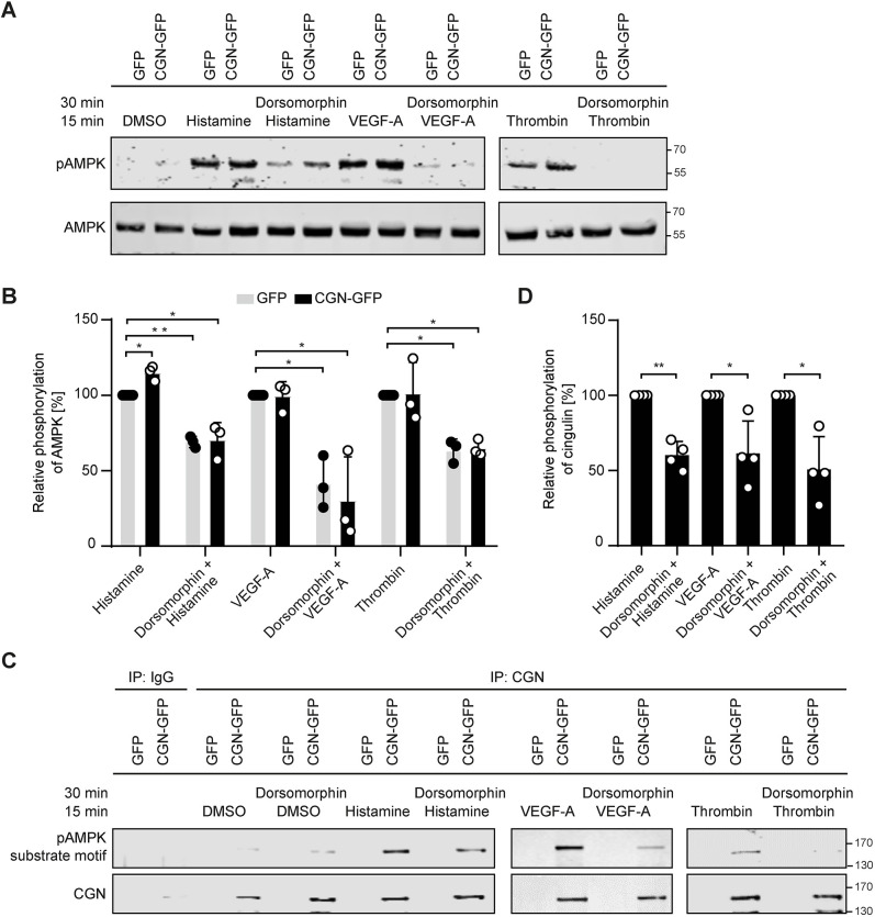
AMPK activation induces cingulin phosphorylation. (A) Representative western blots of cingulin-overexpressing HUVECs (CGN-GFP) and control HUVECs (GFP) stimulated with histamine (10 μg/ml), VEGF-A (50 ng/ml) or thrombin (0.5 U/ml) for 15 min, or pre-treated with the AMPK inhibitor dorsomorphin (10 μM) for 30 min are shown. (B) Quantitative analysis of the western blotting data. AMPK was activated in histamine-, VEGF-A- and thrombin-stimulated cells but was inhibited in dorsomorphin pre-treated cells. No significant difference between cingulin overexpressing HUVECs (CGN-GFP) and control HUVECs (GFP) was found (n=3). *P<0.05, **P<0.01 (one-way ANOVA with Tukey's post-hoc test). (C) Cingulin phosphorylation was determined by recognition of the pAMPK substrate motif (LXRXXS/T). Cingulin phosphorylation was verified using immunoprecipitation (IP) and western blotting. (D) Quantitative densitometry analysis of western blotting data. Cingulin was phosphorylated after stimulation, and phosphorylation was decreased by the AMPK inhibitor dorsomorphin. Data are presented as the mean±s.d. (n=4). *P<0.05, **P<0.01 (one sample t-test).

