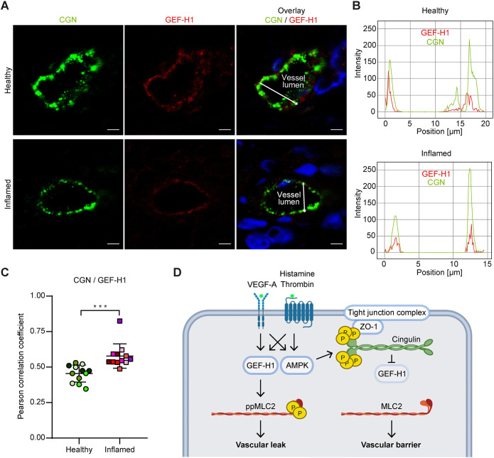
Fig. 8.
Cingulin and GEF-H1 colocalise in inflamed small blood vessels of human skin samples. (A) Healthy and inflamed vessels of human skin tissues were stained for cingulin (green) and GEF-H1 (red). Nuclei were stained with DAPI (blue). Scale bars: 5 μm. (B) Immunofluorescence intensity graphs are shown for cingulin (green) and GEF-H1 (red) for the position denoted by the white arrow. (C) The colocalisation of cingulin and GEF-H1 was quantified based on the corresponding correlation coefficients. Datapoints for the same patient are denoted by use of the same colour (n=12). Data are presented as the mean±s.d. ***P≤0.001 (Student's t-test). (D) Schematic diagram of the proposed mechanism of the protective effect of cingulin (created using BioRender.com). AMPK-mediated phosphorylation of cingulin regulates its colocalisation with GEF-H1. Binding between GEF-H1 and cingulin leads to decreased MLC2 phosphorylation and reduced vascular permeability.

