. 2021 Sep 3;10:e68542. doi: 10.7554/eLife.68542

Figure 6. Structural analysis of the heparin-binding motif (HBM) in the 3D models for the R568S, R571T, and R568S/R571T mutants compared with the crystal structure of hDAO-WT.

(A) Top view of the HBM in hDAO-WT. R568 and R571 (green sticks) are the key residues in the positively charged HBM formed by arginines and lysines from both chains in the hDAO-WT dimer. Intramolecular interactions formed by R571 stabilize the HBM. (B) Top view of the HBM in hDAO-R568S dimer. The positively charged area is reduced around S568s in the R568S mutant compared to hDAO-WT (A). (C) Top view of the HBM in hDAO-R571T. The stabilizing interactions formed by R571 in hDAO-WT (A) are lost, and positive patches around T571s are reduced in hDAO-R571T. (D) Complex of hDAO-WT dimer with heparin hexasaccharide, sliced side view. R568s form hydrogen bonds with heparin hexasaccharide. (E) Complex of hDAO-R568S/R571T dimer with heparin hexasaccharide, sliced side view. In the R568S/R571T mutant, the positively charged surface patches and the stabilizing interactions are lost. Red color corresponds to the negatively charged surface, blue color indicates positively charged regions. hDAO: human diamine oxidase.

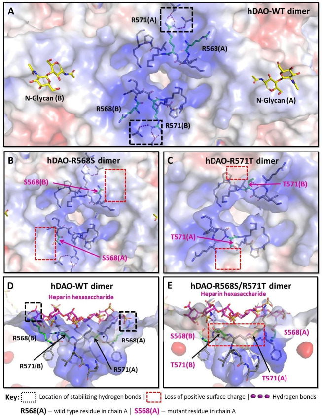
Figure 6.

Figure 6—figure supplement 1. Structural analysis of the heparin-binding motif (HBM) in 3D models of the K575T, K570G/R571Q/K572T, and R568S/R571T mutants versus the crystal structure of hDAO-WT.

Figure 6—figure supplement 1.

(A) HBM in hDAO-WT, top view. (B) Complex of hDAO-WT (left) and hDAO-K575T (right) dimers with heparin hexasaccharide, sliced side view. Change of lysine to threonine leads to a dramatic decrease in the positively charged surface inside the HBM. (C) Local interaction networks of K575 (chain A) in hDAO-WT and the corresponding site with T575 (chain B) of hDAO-K575T show that the mutation eliminates stabilizing hydrogen bonds. (D) Local interaction network of R568 is shown in chain A and R571 – in chain B of hDAO-WT. R568 interacts with heparin hexasaccharide, and R571 establishes multiple interactions with other residues stabilizing the HBM. (E) Local interaction networks are shown for S568 in chain A and T571 – in chain B of hDAO-R568S/R571T. The double mutation leads to an evident disruption of the hydrogen bonding network due to R571T mutation and loss of interactions with the heparin hexasaccharide due to R568S mutation. (F) Local interaction networks of K570, R571, and K572 (chain A) in hDAO-WT compared to the corresponding site in hDAO-K570G/R571Q/K572T (chain B). The triple mutation reduces the number of favorable interactions with other binding site residues and heparin hexasaccharide. (G) Top view of hDAO-WT compared to the K570G/R571Q/K572T triple mutant (chain B). The surface charge of the HBM changes from positive to negative charge in the triple mutant. Red color corresponds to the negatively charged surface, blue color indicates positively charged regions. hDAO: human diamine oxidase.
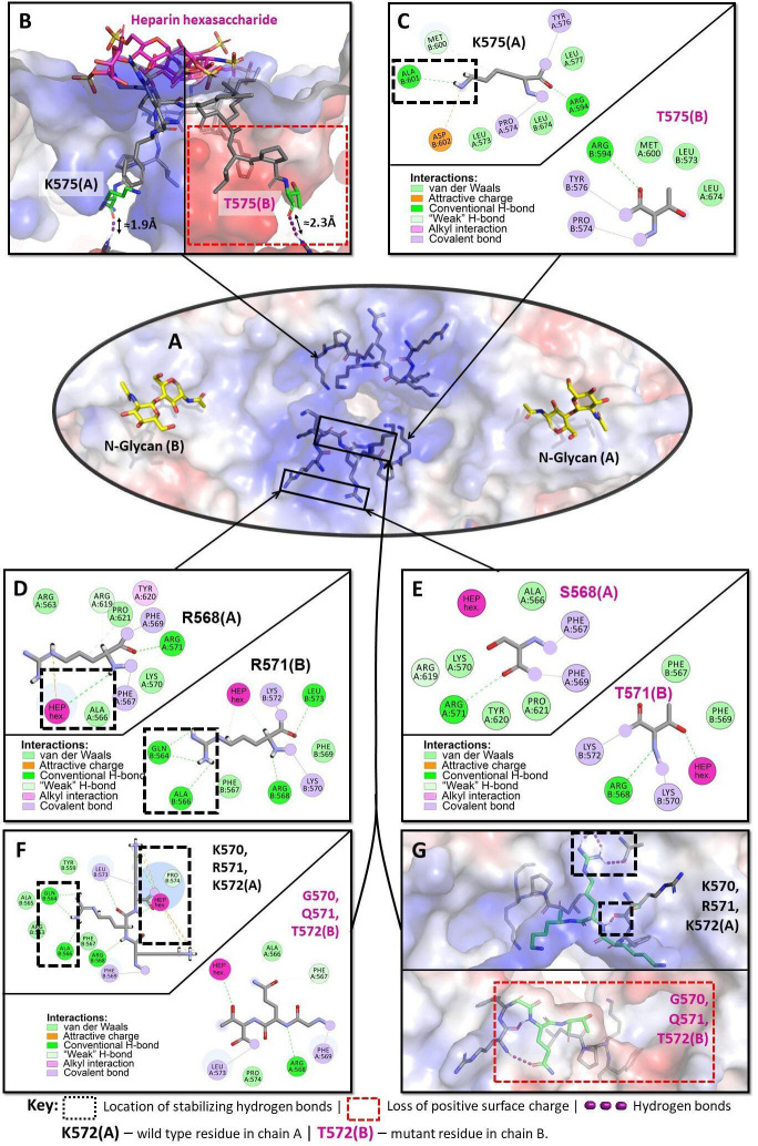
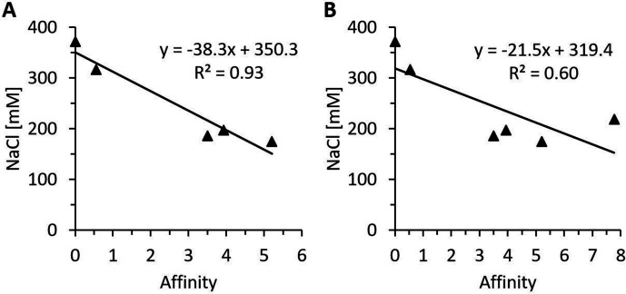
Figure 6—figure supplement 2. Regression analysis of in silico affinity change estimations and measured heparin-sepharose salt elution concentrations of various heparin-binding motif (HBM) mutants.

Figure 6—figure supplement 2.

Regression analysis of predicted free energy changes in heparin affinity and the NaCl concentrations necessary for the elution from heparin-sepharose of the HBM mutants rhDAO-R568S, rhDAO-K575T, rhDAO-R568S/R571T, and rhDAO-R568S/K575T versus rhDAO-WT (0 affinity; 372 mM NaCl). The rhDAO-K570G/R571Q/K572T triple mutant corresponding to the HBM in guinea pig, dog, rat, mouse, and Chinese hamster proteins was excluded (A, p-value 0.0088), or included (B, p-value 0.08) in the calculations.