. 2021 Sep 2;10:e65528. doi: 10.7554/eLife.65528

Figure 6. Generation of hematopoietic stem and progenitor cell-derived plasmacytoid dendritic cells from circulating HSPCs (cHSPCs) from peripheral whole blood using optimized current good manufacturing process (cGMP)-compliant medium.

cHSPCs were pre-expanded for 4 days at low density (1–5 × 10⁵ cells/mL) in cGMP-compliant medium (SCGM) supplemented with UM171 and then cryopreserved. Subsequently, cells were thawed, phenotyped for CD34, and 1 × 10⁵ cHSPCs were seeded for HSPC-pDC generation. HSPC-pDCs were isolated after 16 days of culture and phenotypically analyzed. (a) Calculated number of cells during HSPC-pDC differentiation using pre-expanded HSPCs (without the pre-expansion factor taken into account). (b) Calculated number of HSPC-pDCs upon isolation of HSPC-pDCs at 16 days of culture (with fold pre-expansion taken into account). (c) Percentage of HSPC-pDCs of the total population of cells. (d, e) Levels of type I IFN upon stimulation of HSPC-pDCs with the TLR7 agonist R837 (d) or the TLR9 agonist CpG-2216 (e). Data shown represent ± SEM of four donors (a–c) and four donors each analyzed in technical triplicates (d, e).

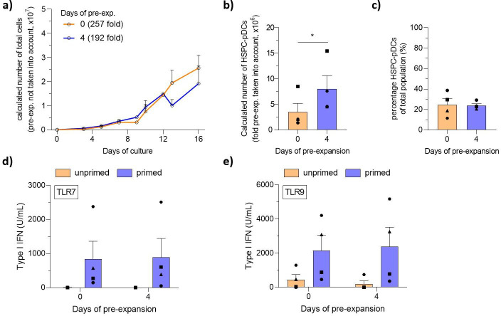
Figure 6—source data 1. Source data related to Figure 6a-e.

Figure 6.

Figure 6—figure supplement 1. Differentiation of circulating CD34+ hematopoietic stem and progenitor cell-derived plasmacytoid dendritic cells (cHSPC-pDCs) from cHSPCs using optimized current good manufacturing process (cGMP)-compliant conditions.

Figure 6—figure supplement 1.

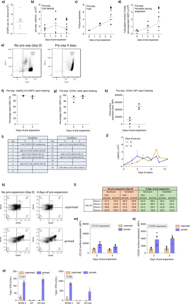
Circulating CD34+ HSPCs (cHSPCs) were isolated from buffy coats and cells were either directly cryopreserved or pre-expanded at low density (1–5 × 10⁵ cells/mL) for 4 days and then cryopreserved. Cells were then thawed, phenotyped, and 1 × 10⁵ HSPCs were seeded for HSPC-pDC generation. (a) Numbers of cHSPCs that can be isolated per mL of blood (assuming 450 mL blood per buffy coat) for eight healthy donors. (b) HSPC density during pre-expansion prior to medium change. Arrows indicate time points when HSPCs were cryopreserved. (c) Fold pre-expansion of cHSPCs. (d) Calculated number of pre-expanded cHSPCs. (e) Representative flow cytometry plots showing the expression of CD34 on freshly isolated cHSPCs or cHSPCs after pre-expansion for 4 days. (f) Viability of cells. (g) Percentage of CD34+ cells. (h) CD34 surface expression of cells. (i) Table showing when cells were split during differentiation of cHSPCs into cHSPC-pDCs. (j) Density of cells during cHSPC-pDC differentiation prior to medium change. (k) Representative flow cytometry plots showing expression of CD123 versus CD303 on lin-CD11c- cells for HSPC-pDCs from the different culture conditions. (l) Table showing the percentage of isolated HSPC-pDCs expressing CD123 and CD303. (m) Surface expression of CD123 on primed or non-primed HSPC-pDCs. (n) Surface expression of CD303 on primed or non-primed HSPC-pDCs. (o) Type I interferon (IFN) response of HSPC-pDCs generated from cHSPCs using either SFEM II medium, DC medium, or DC medium supplemented with ascorbic acid (AA). HSPC-pDCs were activated with either the TLR7 agonist R837 or the TLR9 agonist CpG-2216. Data shown represent ± SEM of eight (a) and four (b–n) donors, and one donor analyzed in technical triplicates (o).
Figure 6—figure supplement 1—source data 1. Source data related to Figure 6—figure supplement 1a-d, f-j, m-o.