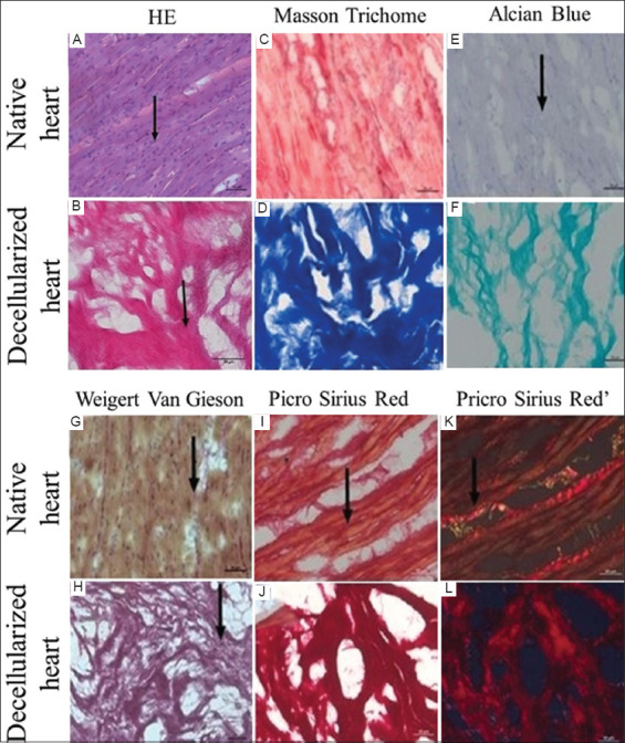
Figure 3. Microscopic analysis of native and decellularized porcine hearts. (A) Native heart – Cardiomyocyte nucleus shown in an arrow, stained by hematoxylin and eosin; (B) Decellularized heart – collagen shown in an arrow, stained by hematoxylin and eosin; (C) Native heart – staining of collagen fibers (dark red) and cardiomyocytes (reddish-pink), stained by Masson’s trichrome; (D) Decellularized heart – collagen fibers highlighted in blue stained by Masson’s trichrome; (E) Native heart – discrete tissue staining, arrow showing discrete area of GAGs, stained by Alcian Blue; (F) Decellularized heart – areas with evidenced GAGs highlighted in greenish-blue, stained by Alcian Blue; (G) Native heart – arrow pointing to a discrete area highlighted in dark red, corresponding to the collagen fiber, interspersed with cardiomyocytes, stained by Weigert-Van Gieson; (H) Decellularized heart – collagen fibers evidenced in dark red, stained by Weigert-Van Gieson; (I) Native heart – collagen fiber highlighted in dark red indicated by the arrow, peripheral to cardiomyocytes, stained with Picrosirius red; (J) Decellularized heart – collagen fibers intensely stained in dark red, stained by Picrosirius red; (K) Native heart – polarized light microscopy highlighting collagen fibers in bright red, stained by Picrosirius red; (L) Decellularized heart – polarized light microscopy with intense highlighting of collagen fibers in pale red, stained by Picrosirius red.

