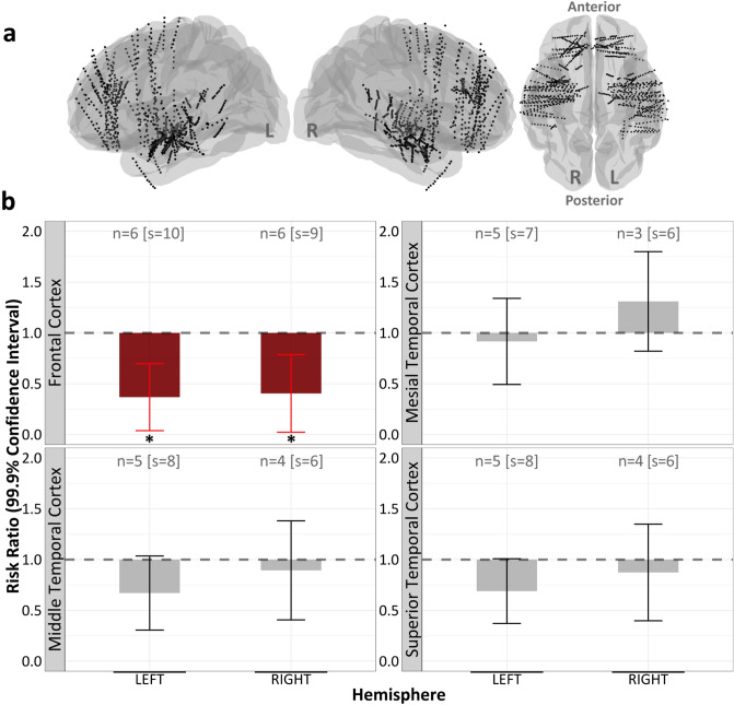
Figure 4.
Bilateral frontal regions responded to K448. (a) Stereo-EEG electrodes aggregated across subjects. (b) Linear mixed models revealed nonsignificant reductions in all regions other than the bilateral frontal cortices (right frontal cortex (FC) % reduction = 59.55, p = 0.049; left FC % reduction = 63.25, p = 0.017). “n” represents the number of subjects with electrode coverage in the corresponding brain region and “s” represents the number of unique experiment sessions. Significance at *p < 0.05, **p < 0.01, ***p < 0.001.

