Figure 1.
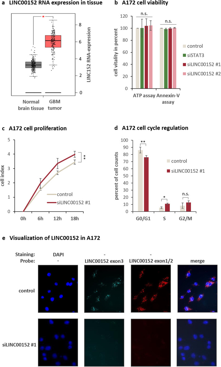
Characterization of LINC00152 functions in A172 glioblastoma cells. (a) GEPIA Plot displayed an increased LINC00152 expression in Glioblastoma Multiforme (GBM) tumor samples compared to healthy donor samples (normal brain tissue). (b) Cell viability was determined 48 h after LINC00152 and STAT3 knockdown using an ATP-based and an Annexin-V assay. Values were normalized to a siRNA negative control. (c) LINC00152 was knocked down and cell proliferation was monitored over time in an incubator equipped with a microscope and camera (XCelligence®). The impedance is recorded by RTCA software (https://www.agilent.com/en/product/cell-analysis/real-time-cell-analysis/rtca-software) and given as cell index, which is a measure of cell proliferation. (d) DNA of A172 cells was labled with propidium iodide 72 h after siLINC00152 #1 and control siRNA transfection and the DNA content was measured by flow cytometry. (e) Specific probes targeting LINC00152 either in exon3 (detecting primary and spliced transcript) or intron-spanning in exon 1 and -2 (detecting just spliced transcripts) were added to formaldehyde fixed A172 cells 48 h after knockdown with siLINC00152 #1 and a negative control siRNA. Nuclei were counterstained with DAPI and images were merged.

