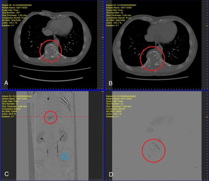
Figure 2.
Screenshot of the image viewer for the observer study. (A) previous computed tomography (CT) image (upper left); (B) current CT image (upper right); (C) projection of temporal subtraction (TS) images (lower left); (D) TS image (lower right). When an observer clicks on a suspicious lesion, the dialog box appears to rate its likelihood (low to high) of being a bone metastasis. These representative images are obtained from a 55-year-old male patient with renal cell carcinoma who developed two osteolytic metastases in a thoracic vertebra (red circle) and the left iliac bone (blue circle). Both metastases are clearly visualized.

