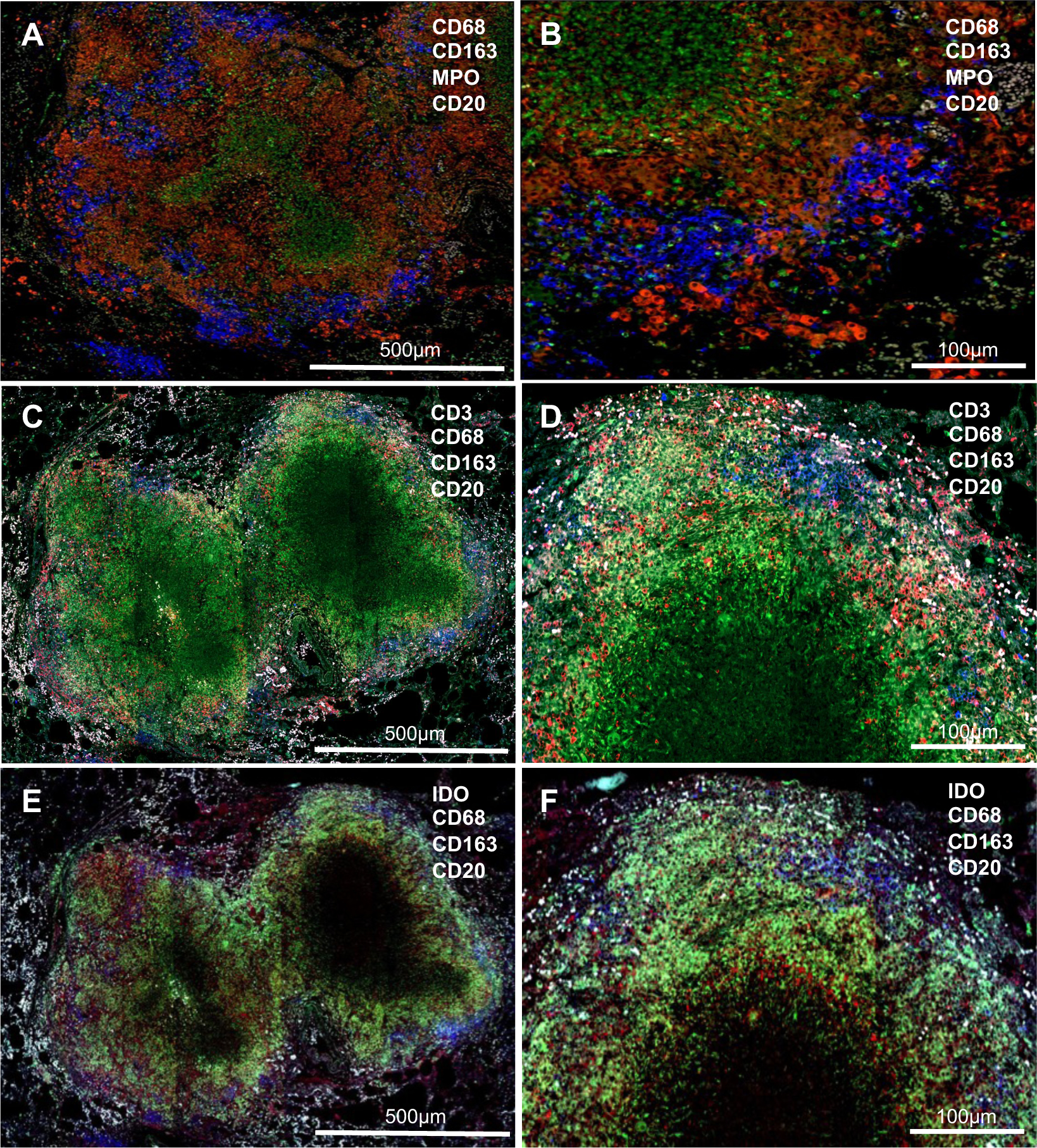
Figure 1. Pulmonary granulomas in infant macaques with tuberculosis.

(A & B) Granulomas, comprising of macrophage layers (red) and clusters of CD20+ B cells (blue), are organized well with a central area of caseous necrosis (MPO-expressing neutrophils); (C-F) Less organized coalescing granulomas, surrounded by a layer of macrophages (green) and IDO-expressing cells (red, E & F) with infiltration of CD3+ T cells (red, C & D). Clustered 20+ B cells (blue) distributed along the layer. MPO, myeloperoxidase; IDO, Indoleamine-pyrrole 2,3-dioxygenase.
