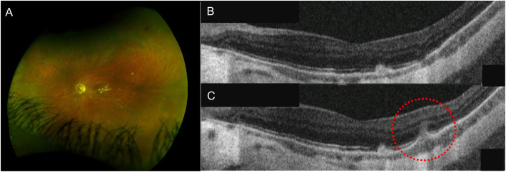
Fig. 4.
Punctate Inner Choroidopathy. A Left eye ultra-wide field fluorescein angiography showing choriorerinal scars located at the posterior pole; B correlating with atrophy of the outer retina and retinal pigment epithelium (RPE) on optical coherence tomography (OCT). C OCT showing elevation of the RPE with underlying hyporeflective space between the RPE and Bruch’s membrane (circle) and increased penetration of light through the inner choroid, suggestive of disease recurrence

