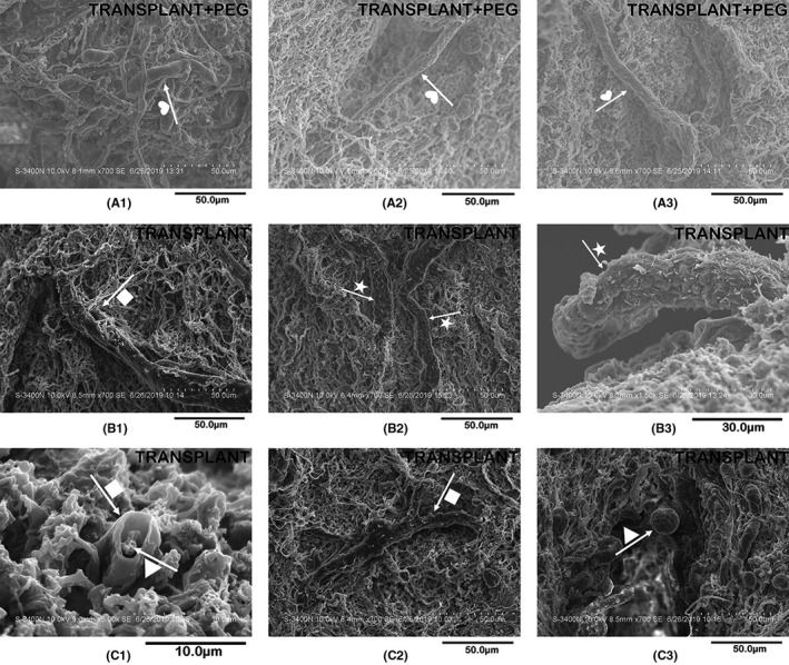
FIGURE 6.

Scanning electron microscopy. In the bridging tissue of the TRANSPLANT group, most of the myelin is damaged (arrow + star in B2 and B3) and incomplete (arrow + square in B1 and C2). By comparison, the myelin of the TRANSPLANT+PEG group has a smooth surface and a complete structure (arrow + heart in A1, A2 and A3). At the edge of the sample blocks in the TRANSPLANT group, we find degenerated myelin (arrow + square in C1) enveloping a degenerated axon (arrow + triangle in C1, similar to the results in Figure 5, namely arrow + heart in A2). In the scars of the TRANSPLANT group, we also find axons with a spherically enlarged stump after degeneration (arrow + triangle in C3)
