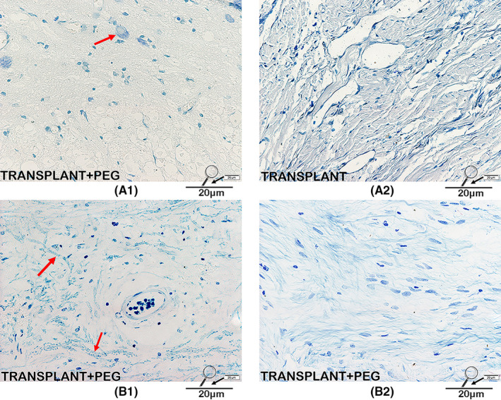
FIGURE 8.

Nissl/LFB staining. Neurons labeled with Nissl bodies can be seen in the bridging tissue of the TRANSPLANT+PEG group (red arrow in A1) but not in the TRANSPLANT group (A2). On LFB staining, irregularly arranged myelin can be seen in the bridging tissue of the TRANSPLANT+PEG group (red arrows in B1), suggesting regrowth, while in the TRANSPLANT group, only stained myelin debris can be seen (B2)
