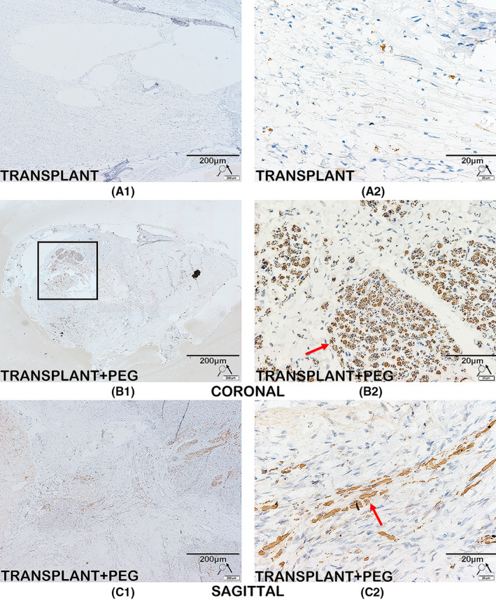
FIGURE 10.

Immunohistochemistry for neurofilament protein. Immunolabeling of neurofilament protein is seen in the TRANSPLANT+PEG group (B1, C1 and red arrows in B2, C2), but not in the TRANSPLANT group (A1 and A2). We also find areas with high concentrations of axons in coronal slices (black square in B1); this is a sign of the survival of the transplanted spinal cord
