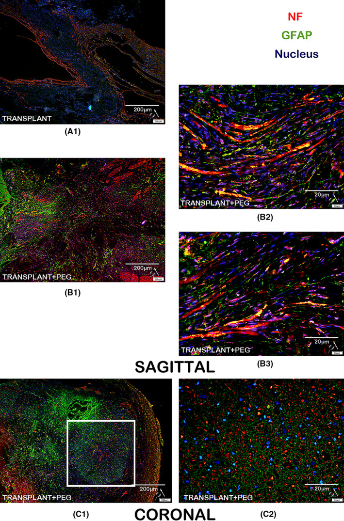
FIGURE 12.

Immunofluorescence for neurofilament protein and GFAP: in the bridging tissue of the TRANSPLANT+PEG group, we observe a large amount of NF immunofluorescence indicative of neuronal fibers (B1 and C1), but we did not find similar immunofluorescence in the bridging tissue of the TRANSPLANT group (A1). In the magnified view, many apparent axons penetrate through the bridging tissue (B2, B3 and C2). In the coronal view, there is dense myelin (white square in C1)
