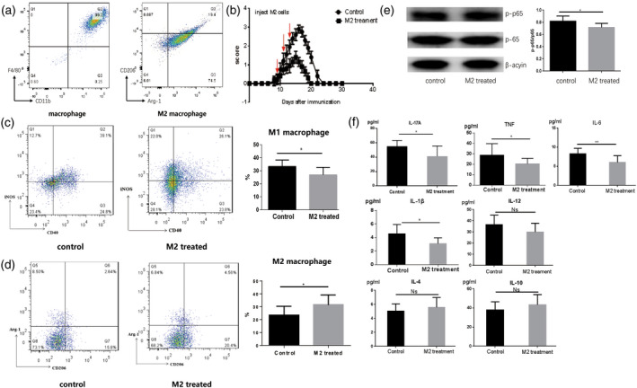
FIGURE 2.

M2 macrophage‐treated experimental autoimmune neuritis (EAN). (a) Macrophage colony‐stimulating factor (M‐CSF) and interleukin (IL‐4)‐induced M2 macrophages. On the 8th day after in‐vitro culture, we evaluated the proportions of macrophages and M2 macrophages. According to flow cytometric analysis, the proportions of macrophages and M2 macrophages were 98.48 ± 0.189% and 94.60 ± 1.407%, respectively. (b) M2 macrophages treatment ameliorated the severity of EAN. We administered M2 macrophages (0.1 ml, 1 × 10⁶ cells) to EAN rats via the caudal vein from days 8 to 14. The scores for the severity of EAN decreased in the M2‐treated group compared to rats in the control group (p < 0.05). Notably, in the M2‐treated group, the duration of clinical symptoms was shorter than the control group. (c–d) The percentage of M1 macrophages was reduced and the percentage of M2 macrophages was increased in spleen mononuclear cells (MNCs) of the M2 macrophage‐treated group compared to the control group. (e) M2 macrophage treatment reduced the level of p‐p65 in sciatic nerves of EAN mice during the peak phase when compared to the control group. (f) The levels of interleukin (IL)‐17A, IL‐1β, IL‐6 and tumor necrosis factor (TNF)‐α decreased greatly compared to the control group. No marked differences appeared for the expression of IL‐12, IL‐4 and IL‐10 among the two groups, but the levels of IL‐4 and IL‐10 increased, although there was no significance
