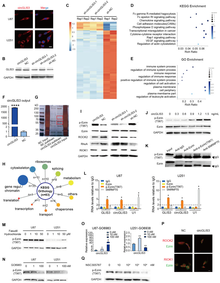
FIGURE 3.
CircGLIS3 directly binds with p-Ezrin(T567) and elevates the p-Ezrin(T567) level in glioma. (A) Fluorescence in situ hybridization (FISH) assay with Cy3-labeled circGLIS3 probe (magnification, ×640; scale bar, 10 μm). (B) Western blot of GLIS3 protein after the upregulated or downregulated circGLIS3 level in U251 cells. (C) RNA-Seq analysis of U251 cells transfected by sh-circGLIS3 or sh-NC lentivirus. (D) KEGG and (E) GO enrichment analysis in U251 cells. (F) RNA pull-down experiments with U251 extract. Pull-down probe efficiency was determined by RT-PCR. (G) RNA pull-down experiments with U251 extract. Specific bands were identified by mass spectrometer. (H) Summary of circGLIS3 binding proteins according to KEGG. (I) Western blot detection after upregulated or downregulated circGLIS3 in U251. (J) Western blot detection of p-Ezrin(T567) in U87 transfected by concentration gradient circGLIS3 plasmid. (K) Western blot of RIP lysate by anti-Ezrin, anti-p-Ezrin(T567), anti-IgG, and anti-SNRNP70 antibodies. (L) RT-PCR analysis of Ezrin, p-Ezrin(T567), IgG-negative control, and SNRNP70-positive control immunoprecipitated RNA of U87 (left) and U251 (right). (M) Western blot of p-Ezrin(T567) in U87 and U251 after being treated with DMSO or fasudil hydrochloride for 2 days. (N) Western blot of U87 and U251 after treated with GO6983 for 2 days. (O) RT-PCR of U87 and U251 after being treated with GO6983 for 2 days. (P) Confocal microscope image of Ezrin and ROCK2 or RIOK1. Scale bar, 5 μm. (Q) Western blot of p-Ezrin(T567) in U87 after being treated with NSC305787 for 9 h. The results are presented as the mean ± SEM of biological triplicate assays. *P < 0.05, **P < 0.01, ****P < 0.0001.

