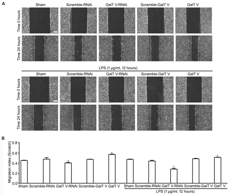
FIGURE 3.
Acceleration of β-1, 4-GalT V on wound healing. HAPI cells with designated constructions as indicated were treated with LPS (1 μg/ml, 24 h). (A) Representative images for wound recovery; scale bar, 100 μm. (B) Quantification of wounded area [migration index (scratch) = wounded size after 24 h/initial wound area)]. Groups significantly different from the non-treated sham group were marked by asterisks. Graphs show mean ± SD; n = 3; *p < 0.05.

