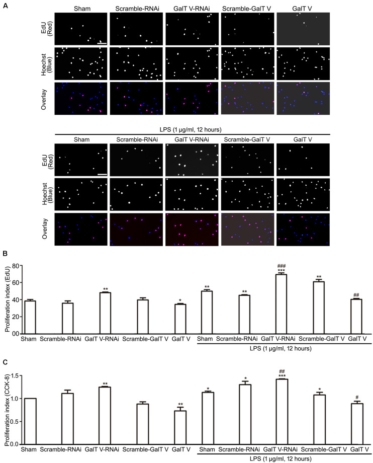
FIGURE 4.
Inhibition of β-1, 4-GalT V on cell proliferation. HAPI cells with designated constructions were treated with LPS (1 μg/ml, 12 h), as indicated. Proliferation of HAPA cells was detected by EdU assay (A,B) and CCK-8 test (C). (A) Representative images for DNA replicating cells by red fluorescence in nucleus; scale bar, 100 μm. (B) Quantification of DNA replicating cells [proliferation index (EdU) = DNA replicating cells/Hoechst positive cells × 100%]. (C) Quantification of cell proliferation by CCK-8 test [proliferation index (CCK-8) = absorbance/absorbance in non-treated sham group]. Groups significantly different from non-treated sham group, LPS-treated sham group were marked by asterisks, hashtags respectively. Graphs show mean ± SD; n = 3; *p < 0.05, **p < 0.01, ***p < 0.001, #p < 0.05, ##p < 0.01, ###p < 0.001.

