
Figure 1.
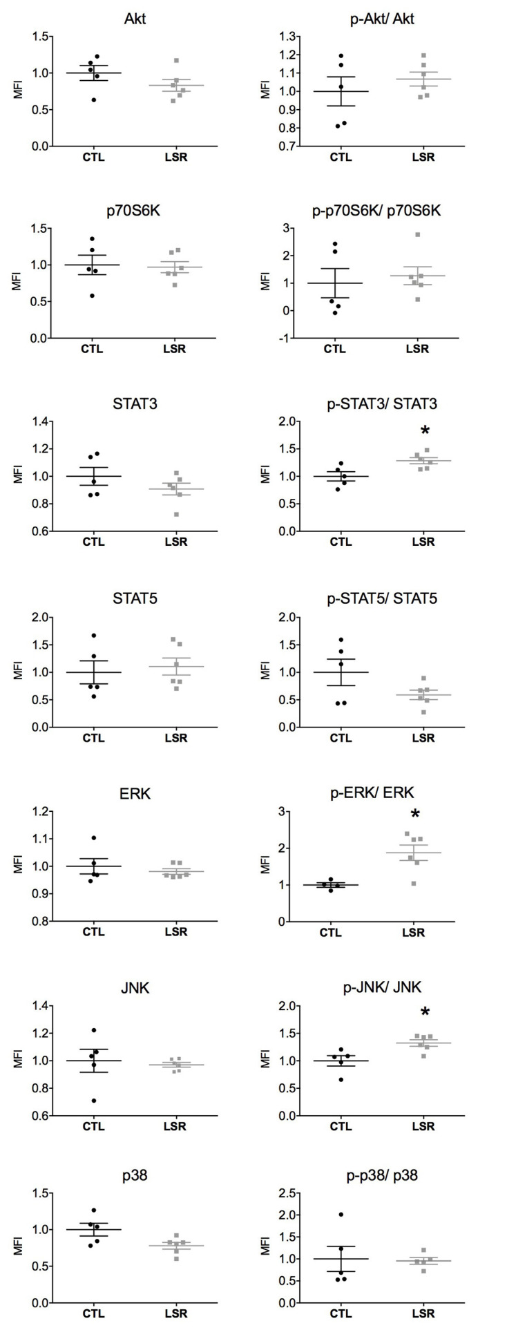
Cortical expression and activation (phosphorylated/total protein) of intracellular signaling pathways in rats from the laser (LSR) and control (CTL) groups. Data are presented as individual points to show dispersion and as mean and standard error of the mean (±SEM). Significant increases in the activation of STAT3, ERK, and JNK were found in the laser group when compared to the control group (*). Data were normalized to the mean fluorescence intensity (MFI) of the control group (p < 0.05; Mann-Whitney U-Test).