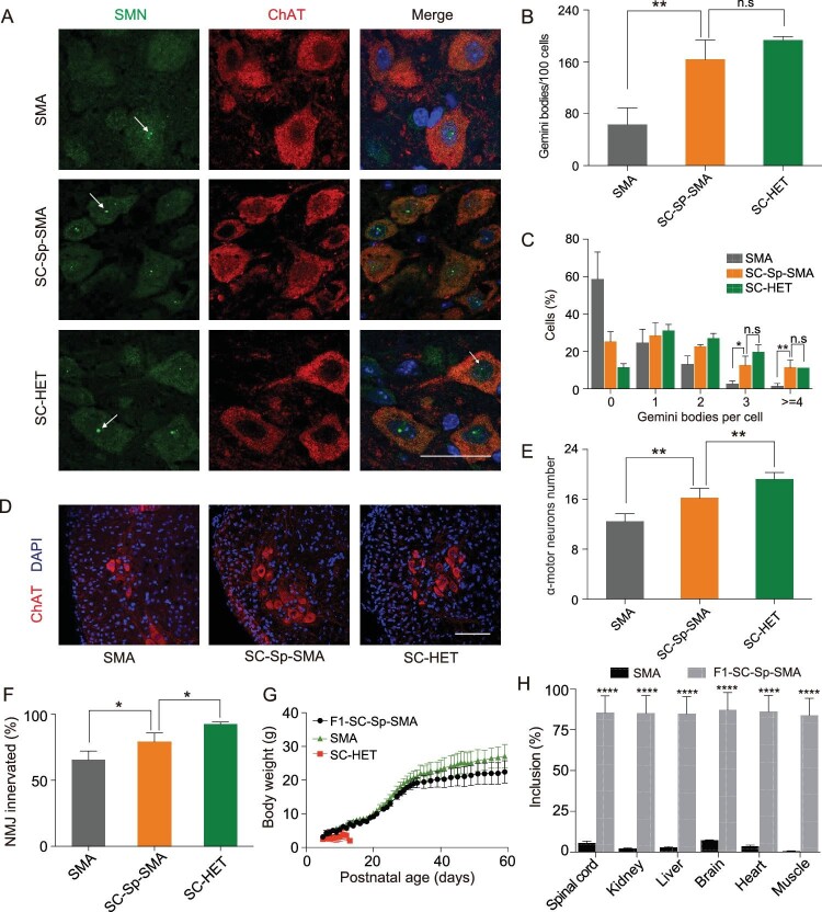
Figure 3.
SMN restoration alleviates spinal MN degeneration and NMJ denervation in SC-Sp-SMA mice. (A) Nuclear Gemini bodies localization in spinal-cord L1–L2 motor neurons was determined by co-staining with SMN and ChAT antibodies. Nuclei were counterstained with DAPI. Gemini bodies are indicated by arrows. Scale bar, 50 μm. (B) and (C) Quantification of Gemini bodies per 100 motor neurons (B) and percentage of motor neurons containing zero, one, two, three, four or more Gemini bodies (C) in SMA (n = 3), SC-Sp-SMA (n = 6) and SC-HET (n = 3) mice. (D) Representative images of spinal-cord L1–L2 ventral-horn ChAT+ MNs (red) in the above three groups. Scale bar, 100 μm. (E) MNs labeled in (D) were counted. SMA (n = 6), SC-Sp-SMA (n = 5) and SC-HET (n = 6). (F) Quantification of the innervated NMJs in SMA (n = 3), SC-Sp-SMA (n = 5) and SC-HET (n = 4) mice. (G) Body weight was assessed from P5 to P59 for F1-SC-Sp-SMA (n = 9). Data from SMA mice and SC-HET were the same as in Figure 2 (I). (H) Statistical analysis of SMN2 mRNA in multiple tissues from SMA and F1-SC-Sp-SMA mice. Error bars indicate means ± SDs. ns, not significant; *P < 0.05; **P < 0.01; ****P < 0.0001; one-way ANOVA.

