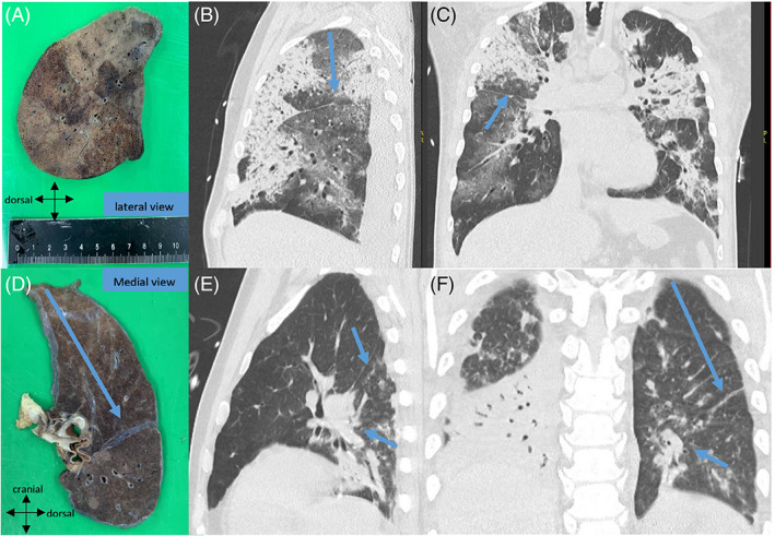
FIGURE 1.

Fresh resection specimen of right lung photographed from the lateral view (A). Distance from the lobar apex and fissure relevant to the CT‐pattern of interest is measured on the CT scan and subsequently in the specimen in order to reach exactly the same level (B,C). (D) Another example of the left lung photographed from the medial view which correlates with the premortem CT image (E,F) in order to correlate with the oblique fissure and bronchial branch (arrows)
