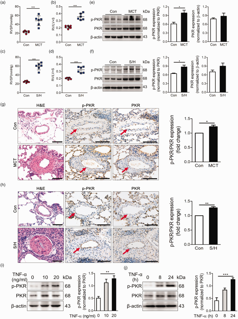
Fig. 1.
PKR activation is elevated in the pulmonary vessels of experimental PH models. (a, b) Right ventricle systolic pressure (RVSP) (a) and indices of RV weight (RV/LV+S) (b) in rats given vehicle or MCT (60 mg/kg, i.p.) (n = 7). (c, d) Right ventricle systolic pressure (RVSP) measurements (c), indices of RV weight (RV/LV+S) (d) in vehicle and SU5416/hypoxia rats (n = 6). (e) Representative immunoblots and densitometric analysis of pulmonary artery PKR and p-PKR level in control and MCT rats (n = 3). (f) Representative immunoblots and densitometric analysis of pulmonary artery PKR and p-PKR level in control and SU5416/hypoxia rats (n = 3). (g) Photomicrographs of serial sections of peripheral rat lung containing small arteries from control animals and rats exposed to MCT rats. Sections were immunostained for the hematoxylin-eosin, PKR and p-PKR. Scale Bar = 25 µm. (h) Photomicrographs of serial sections of peripheral rat lung containing small arteries from control animals and SU5416/hypoxia rats. Sections were immunostained for the hematoxylin-eosin, PKR and p-PKR. Scale bar = 25 µm. (i) Representative immunoblots and densitometric analysis of PKR activation level was assessed in PAECs with TNF-α at 0, 10 and 20 ng/ml (n = 4). (j) Representative immunoblots and densitometric analysis of PKR activation level in PAECs under the treatment of TNF-α (20 ng/ml) for 0, 8 and 24 h (n = 4). *P < 0.05, **P < 0.01, ***P < 0.001, by one-way ANOVA relative to control groups.

