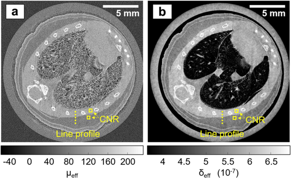
Fig. 8.
Sub-pixel resolution EI XPC micro-CT images of a mouse chest, acquired with the dithered, multi-franie scheme. (a) Reconstructed attenuation image. (b) Reconstructed phase image. Figure reprodueed from [79] without modifieation under the Creative Commous CC BY license. Note that the line profiles and CNR boxes shown in this figure were used to measure contrast-to-noise ratios and spatial resolution in the cited work.

