Figure 2.
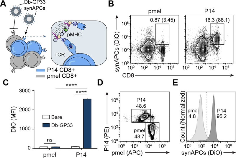
Synthetic APCs bind to T cells in an antigen-specific manner. (A) Db-GP33 synAPCs are incubated with CD8+ T cells from P14 and control pmel TCR transgenic mice. (B) Flow plots of synAPC binding to CD8+ T cells from pmel or P14 TCR transgenic mice. Frequencies depicted are based on gating on total splenocytes and gating on CD8+ cells. (C) MFI of CD8+ cells from pmel or P14 transgenic mice stained with untargeted or Db-GP33 synAPCs. Data shown as mean ± s.d. n=3 ****p<0.0001 by two-way ANOVA with Tukey’s multiple comparison test. (D) Flow plot of pmel and P14 CD8+ T cell mixture used in competition assay. pmel and P14 cells were pre-stained with αCD8-APC and αCD8-PE, respectively, prior to combining. (E) Flow plot of competition assay in which a mixture of pmel and P14 CD8+ T cells are incubated with Db-GP33 synAPCs.

