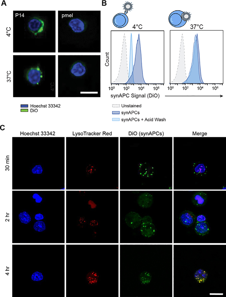
Figure 4.
Synthetic APCs are rapidly internalized by antigen-specific T cells. (A) Representative images of P14 and pmel CD8+ T cells stained with DiO-labeled Db-GP33 synAPCs at 4 or 37°C. Scale bar = 15 μm. (B) Representative histograms for cells stained with synAPCs at 4 or 37°C and analyzed by flow cytometry before and after treatment with an acid wash to strip cell surface receptors. (C) Representative images of CD8+ T cells labeled with a lysosome dye and stained with synAPCs for various lengths of time. Cells were fixed at the times indicated and imaged by confocal microscopy. Scale bar = 15 μm.

