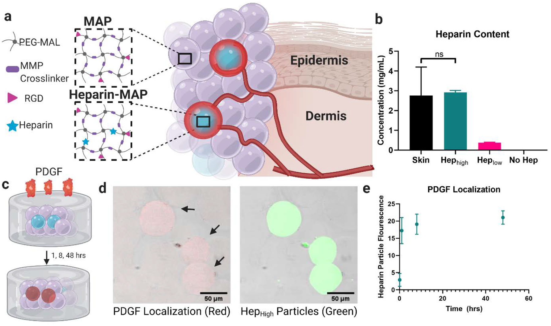
Figure 1.

Heparin μIsland Composition and Growth Factor Sequestration. a) Particles were composed of a PEG-maleimide backbone and MMP-2 cleavable crosslinker with an RGD cell adhesive peptide with or without thiolated heparin. Small fractions of heparin particles (μIslands) were mixed with no-heparin particles to generate growth factor depots. b) Heparin concentration within the particles was matched to mouse skin (HepHigh) and one tenth of mouse skin (HepLow). c) To test for PDGF localization around our heparin μislands, we used a well-based assay with heterogenous scaffolds (10% HepHigh) embedded within a collagen-agarose scaffold before introduction of a solution of biotin-labeled PDGF. d) After fixation, fluorescent Streptavidin revealed PDGF localization (red) in the heparin μIslands (green) as early as 1 hour. e) Quantification of fluorescence in the heparin particles across the 48hr time point. Graphs represent mean +/− standard deviation.
