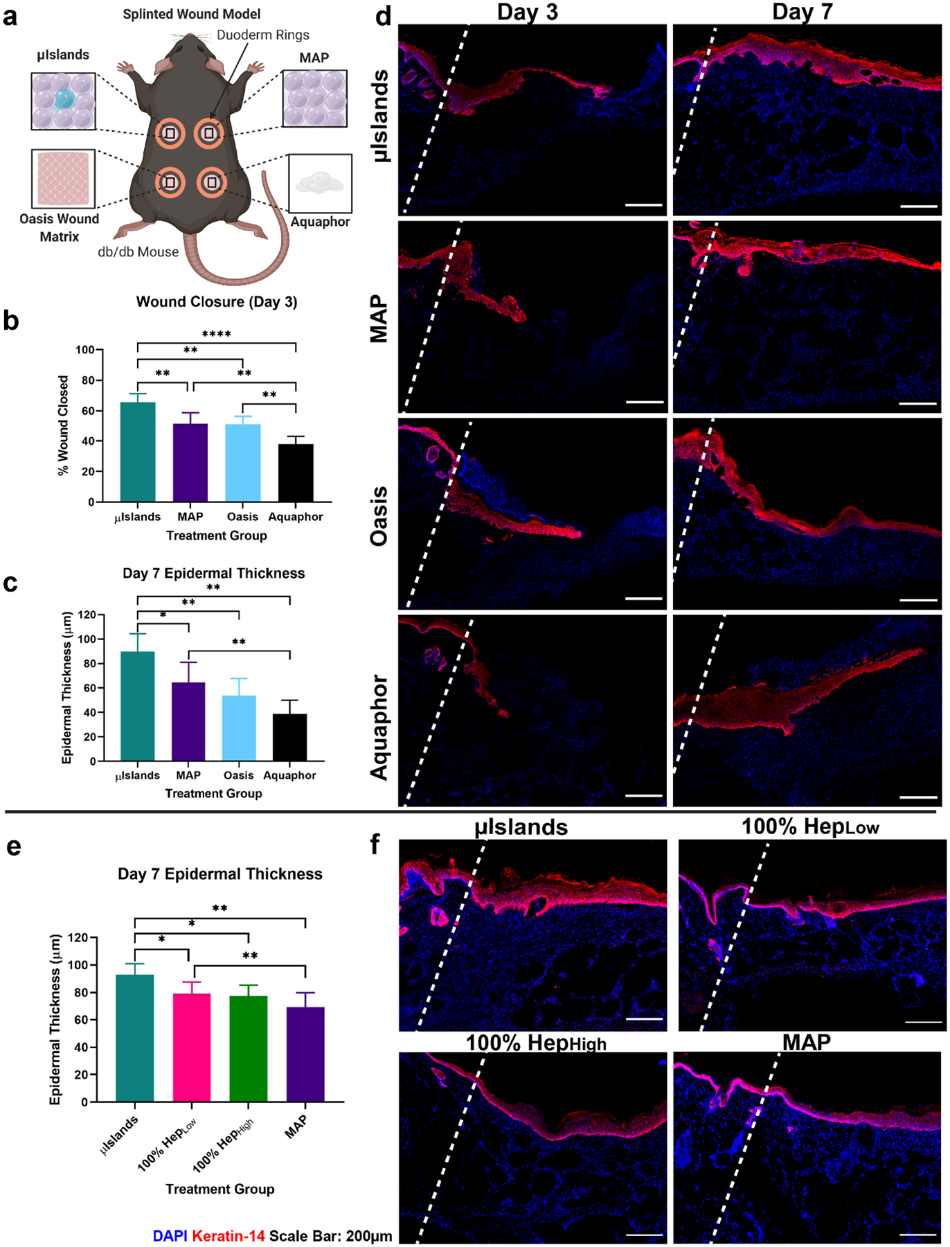
Figure 3.

Epidermal Regeneration in a Diabetic Wound Healing Model. a) Four treatment conditions were evaluated in a mouse diabetic wound healing model at Day 3 and Day 7. b) Wound closure at Day 3 was quantified from the epidermal tongue length. c) Epidermal thickness was quantified for all at Day 7 to compare stages of healing. d) Representative images of the four treatment groups keratin-14 staining at Day 3 and Day 7. e) Day 7 epidermal thickness quantification in a separate study confirmed heparin heterogeneity was necessary for improved epidermal thickness. f) Representative keratin-14 staining from the follow-up study at Day 7. Wound edges marked by dashed line. All graphs show mean +/− standard deviation. Statistics: ANOVA, Multiple comparisons post-hoc tests (Tukey HSD). N=6 for a-d, N=7 for e,f. *p<0.05, **p<0.01, ***p<0.001, ****p<0.0001.
