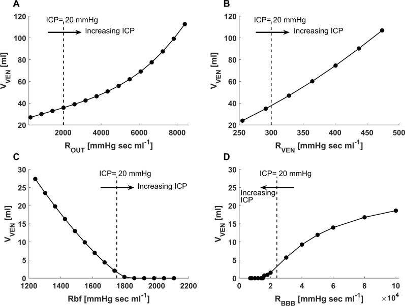
Fig. 6.
Ventricular volume as a function of changes in resistances in the model. A As expected, increased CSF outflow resistance (ROUT) representing non-absorptive hydrocephalus leads to increased ventricular volume (VVEN). B Increased resistance to CSF flow in the ventricular system (RVEN) which depicts obstructive hydrocephalus also leads to increased VVEN. C Conversely, an increase in the resistance to bulk flow of cerebral interstitial fluid (RBF) that occurs with cerebral edema results in a decrease and then collapse of VVEN, simulating the clinical situation in which ventricular volume decreases markedly as the brain swells. D A decreasing resistance to movement of fluid across the blood–brain barrier (RBBB) leads to a rapid decrease in VVEN, simulating the situation in which blood–brain barrier disruption leads to brain swelling with a concomitant decrease in ventricular volume

