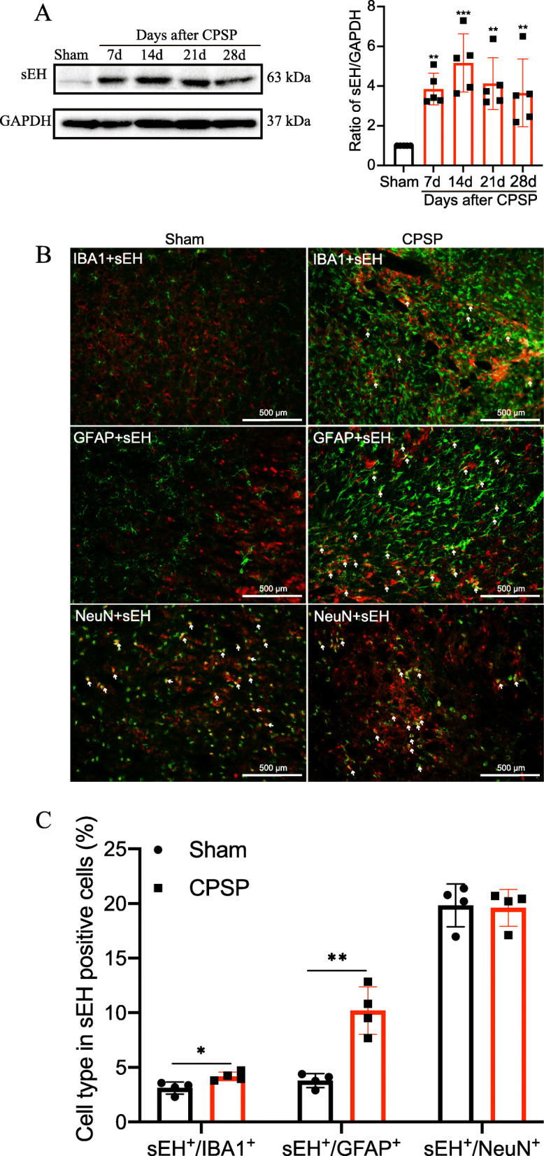
Fig. 2.

Changes of sEH expression in the perithalamic lesion site of CPSP rats. a Representative Western blot bands were presented on the left, with data analysis shown on the right revealing that sEH protein expression is increased around CPSP rat thalamic lesion site, beginning at postlesion day 7 and at least lasting to day 28 after lesion, compared with the sham control. Values are expressed as mean ± SD. GAPDH serves as loading control. **P < 0.01, ***P < 0.001, n = 5 rats per group, one-way ANOVA followed by Bonferroni post hoc test. b The tissue sections from the injured side thalamus are double immunostained with sEH (red) and reactive astrocyte marker GFAP (green), microglial marker IBA1 (green), or neuron-specific nuclei marker NeuN (green). c The percentage of different cell types in sEH-positive cells on day 14 after CPSP induction. n = 4 rats per group, *P < 0.05, **P < 0.01, compared with the sham group. Scale bars = 500 μm. IBA1, ionized calcium-binding adapter molecule 1; NeuN, neuronal nuclei; GFAP, glial fibrillary acidic protein
