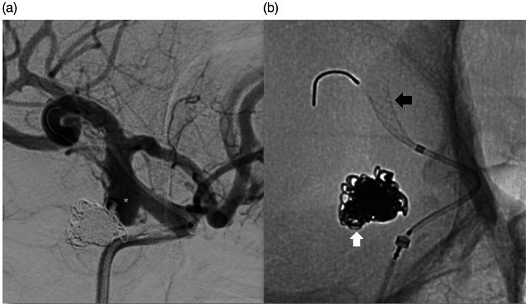
Figure 1.
Angiographic images at the time of attempted flow diverter placement. Digital subtraction angiogram (right anterior oblique projection) of the right internal carotid artery (a) shows the recurrence of the previously coiled right posterior communicating aneurysm (asterisk). Unsubtracted image (b) shows the previous coil mass (white arrow) and the partially deployed flow diverter in the supraclinoid internal carotid artery (black arrow).

