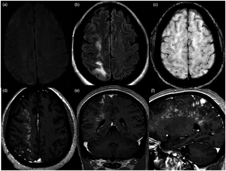
Figure 4.
MRI brain exam performed two days after successful placement of the flow diverters. Axial diffusion weighted images (a) showing no restricted diffusion. Axial FLAIR image (b) shows worsened hyperintensities in the right frontal and parietal subcortical white matter. Axial susceptibility weighted image (c) shows a few associated punctate signal voids and axial (d), coronal (e) and sagittal (f) gadolinium-enhanced T1 images show a dramatic increase in extent of multinodular enhancement.

