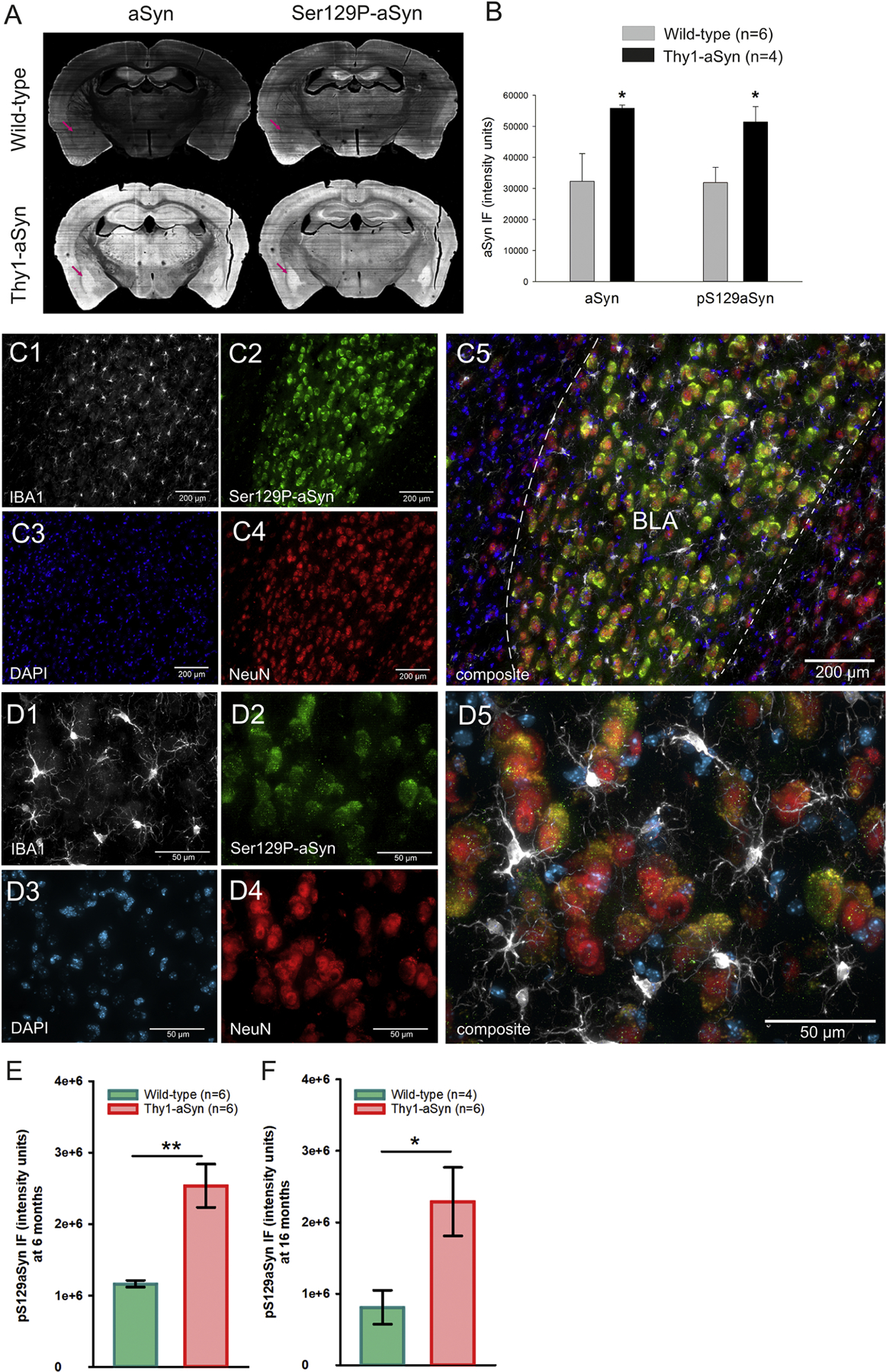
Fig. 6.

Immunofluorescent staining of human and mouse α-syn and phosphorylated α-syn (Ser129P-α-syn) antibodies in sections of the amygdala. A,B) Thy1-aSyn transgenic mice extensively accumulate aSyn and Ser129P-aSyn in the amygdala (purple arrow, mean + SEM, Student’s t-test; *p < 0.05). C, D) Thy1-aSyn transgenic mouse with accumulated Ser129P-aSyn in the BLA at two magnifications. Phosphorylated α-syn accumulation (C2, D2) was apparent in NeuN positive cells (C4, D4), but not all NeuN positive cells contained Ser129P-aSyn; IBA-1 positive cells (C1, D1) and a number of DAPI stained cells (C3, D3) appear devoid of Ser129P-aSyn. IBA-1 positive cells (C1, D1) appear with enlarged cell bodies and activated morphology. Basolateral nucleus of the amygdala (BLA), scale bar = 200 µm (C) or 50 µm (D). E, F Quantification of Ser129P-aSyn accumulation in the BLA of 6 months old (E) and 16 months old (F) mice (mean ± SEM, Student’s t-test; *,**p < 0.05, 0.01, N is provided in figure).
