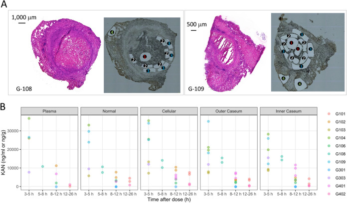
FIG 2.
KAN distribution in human lung lesions. (A) Typical examples of histology staining and laser-capture microdissection (LCM) of thin human lesion sections. Two large necrotic lesions were collected from the resected lung tissue of human subjects G-108 and G-109. Adjacent lesion sections were used for hematoxylin and eosin (H&E) staining (left) to guide LCM sample collection (right); 1, inner caseum; 2, outer caseum; 3, cellular rim; 4, uninvolved lung. Laser-dissected pieces belonging to the same tissue compartment were pooled for quantitation by LC-MS/MS. (B) Concentrations of KAN in plasma and 20 resected human lesions from 10 subjects, collected at various times from 3 h to 26 h after a single dose or multiple doses of 1,000 mg of KAN injected intramuscularly (49). Concentrations were determined in thin section samples collected by LCM and analyzed by LC/MS-MS (47). Each color corresponds to one subject. Raw data are provided in Data Set S1 in the supplemental material.

