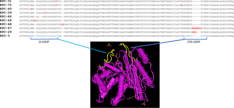
FIG 1.
KPC-3 variants identified in the ceftazidime-resistant Klebsiella pneumoniae strains. Protein sequence alignment of amino acid residue positions 156 to 298 of KPC-3 variants is shown with respect to the KPC-3 reference protein sequence (NCBI reference sequence WP_004152396.1 [59]). Mutation mapping was performed on the MMDB 6QWD crystal structure of KPC-3 downloaded at the NCBI Cn3D macromolecular structure database visualized by the Cn3D 4.3.1 viewer tool (60) at the Cn3D home page (https://www.ncbi.nlm.nih.gov). Amino acid residues involved in the two loops highlighted in yellow in the KPC-3 three-dimensional model are indicated in blue letters. Amino acid substitutions, insertions, and deletions in ceftazidime-avibactam-resistant KPC variants are indicated in red letters.

