
Fig. 7.
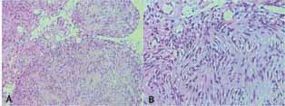
( A, B ) Hematoxylin and eosin–stained sections (low power field [A] and high power field [B] ) showing spindle cells with ill-defined cytoplasm and wavy vesicular nucleus with focal palisading Verocay bodies.
Official websites use .gov
A
.gov website belongs to an official
government organization in the United States.
Secure .gov websites use HTTPS
A lock (
) or https:// means you've safely
connected to the .gov website. Share sensitive
information only on official, secure websites.
( A, B ) Hematoxylin and eosin–stained sections (low power field [A] and high power field [B] ) showing spindle cells with ill-defined cytoplasm and wavy vesicular nucleus with focal palisading Verocay bodies.