
Figure 1.
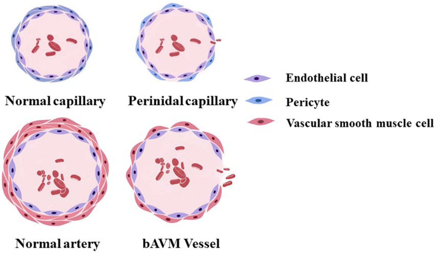
A schematic diagram of a normal capillary, a perinidal capillary, a normal artery and a bAVM vessel. The perinidal capillary has fewer peridytes and the bAVM vessel has fewer vAMCs than normal capillary and artery which render the vessel prone to bleed and rupture.