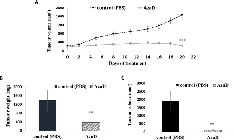
Fig 5. The effect of 5-AzaD on tumour development using xenograft mice.
Athymic nude male mice were SC implanted with 1X10⁶ FaDu cells. When the tumours reached a volume of ~200 mm3 the mice were divided into 2 groups (n = 8). Mice were intraperitoneal (IP) injected three times a week either with PBS X 1 to the control group, or with 2.5 mg/kg of 5-AzaD for 3 weeks. During the experiment, tumour volumes were measured twice a week using calibre meter (A). On day 21 at the end of the experiments, mice were sacrificed, and the tumour tissues were dissected and measured for weight (B) and volume (C). The results were presented as the mean± SD (n = 8). Statistical significance was determined by two tailed student’s–t–test and assigned as **P < 0.01, ***P < 0.001. In order to better understand how 5-AzaD treatment affects tumour growth in vivo, tumours were fixed and sectioned for histological staining. H&E staining indicated that 5-AzaD treatment induced apoptosis of tumour cells and inhibited mitosis when comparing to the control group (Fig 6A). Morphological criteria for diagnosis of mitosis, using the H&E staining, included reporting of increased mitosis above or below the normal background levels, of what is usually seen in control animals [22].

