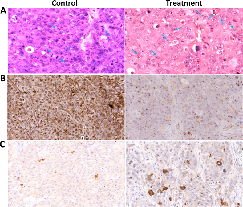
Fig 6. H&E stained images and Ki-67 and active caspase 3 immunostaining of tumour sections from control and treated animals.

Nude mice bearing FaDu tumour cells were treated with 2.5 mg/kg of AzaD or PBS 3 times a week for 3 weeks. At the end of the experiment, mice were sacrificed and the tumour tissues were dissected. (A) Representative H&E stained images of isolated tumours are shown. Mitotic cells in the control and apoptotic cells in the treated groups are marked with arrows. (B) Proliferation profiles of the cells were detected by Ki-67 immunostaining as described under “Material and Methods”. The images are representatives of the results obtained from control and treatment groups (Magnification, 400X). (C) Apoptotic cells were detected by caspase 3 immunostaining positivity; criteria are explained in the materials and methods section. The images are representatives of the results obtained from control and treatment groups.
