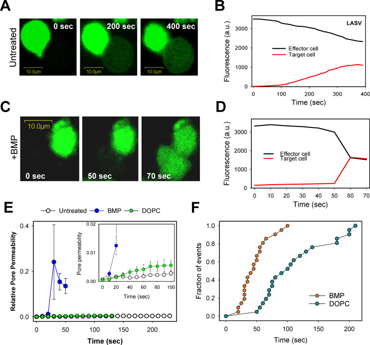
Fig 6. BMP markedly promotes the formation and enlargement of LASV GPC-mediated fusion pores.
Effector COS7 cells expressing GPC were loaded with calcein (green), mixed with unlabeled 293T cells and adhered to poly-lysine coated coverslips. Cells were then pre-treated with 20 μg/ml of BMP or DOPC for 10 min at room temperature or left untreated. Cell-cell fusion was triggered by transferring cells to a pH 5.0 buffer and quickly raising the temperature to 37°C through an IR temperature jump protocol (see Materials and Methods). (A, B) Snapshots and fluorescence intensities showing partial calcein redistribution between untreated effector and target (dye donor and acceptor) cells. (C, D) Same as in panels A, B, but for cells pretreated with BMP. (E) Ensemble average of permeabilities of six fusion pores for untreated and DOPC- or BMP-treated cells. Individual pore permeability traces calculated as described in Materials and Methods were aligned so that t = 0 represents a time point immediately before dye redistribution was detected. Inset: Initial permeability profiles of fusion pores. Error bars are SEM. (F) Kinetics of fusion pore formation for control and BMP-treated cells. The data points represent time after raising the temperature until the onset of calcein transfer.

