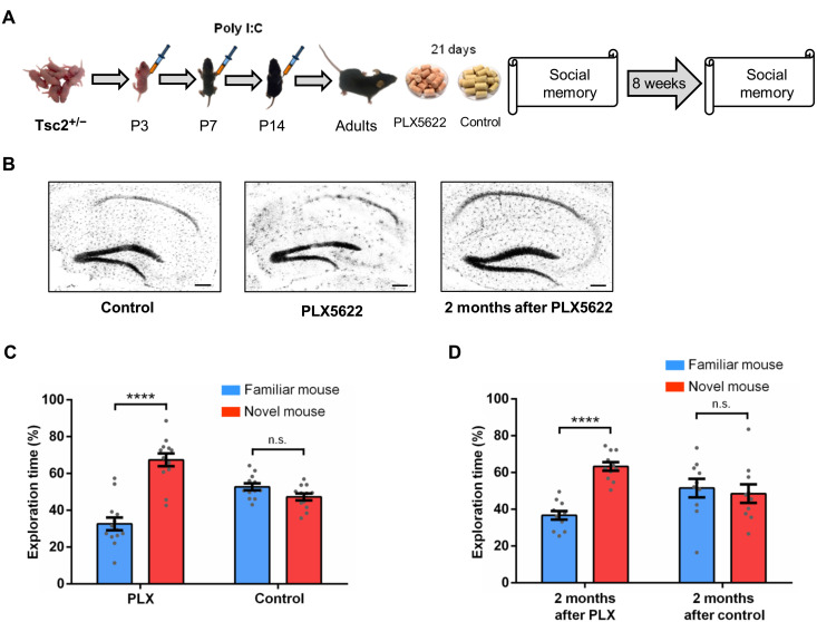
Fig. 3. Effects of microglia depletion in male Tsc2+/− Ep mice.
(A) Timeline for injections of Poly I:C, treatment with PLX5622 (PLX; depletes microglia) or control chow and behavior approach. (B) IBA1 immunostaining of Tsc2+/− control, PLX, and 2 months after PLX mice. The treatment with PLX led to the elimination of most microglia in the whole brain (hippocampus shown as example) compared to the control group (hippocampus shown as example). Two months after PLX, the microglia had repopulated the brain (hippocampus shown as example). (C) Tsc2+/−/PLX mice (n = 13; P < 0.0001, t = 7.10), but not Tsc2+/−/control (n = 12; P = 0.0502, t = 2.07) mice, show normal social memory. (D) Tsc2+/− mice 2 months after PLX (n = 11; P < 0.0001, t = 8.11), but not Tsc2+/− mice 2 months after control (n = 10; P = 0.67, t = 0.42), show normal social memory. Data represent means ± SEM as well as values for individual mice. Scale bars, 200 μm.

