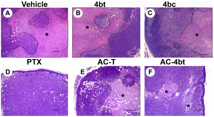
Figure 2. Histopathological analysis of tumor sections upon monotherapy or drug combination treatments.
Representative images of paraffin-embedded sections of tumors stained with H&E from the different experimental groups administered intraperitoneally (i.p.) with: (A) Vehicle, (B) 4bt (80 mg/Kg), (C) 4bc (80 mg/Kg), (D) paclitaxel (20 mg/Kg), (E) Doxorubicin (10 mg/Kg) + Cyclophosphamide (100 mg/Kg) followed by paclitaxel (10 mg/Kg) (AC-T) and (F) Doxorubicin (10 mg/Kg) + Cyclophosphamide (100 mg/Kg) followed by KIF11 inhibitor 4bt (AC-4bt). Treatment with 4bt used as monotherapy or in drug combination prevent exacerbated tumor growth leading to a decrease in the extension of the central necrotic area (outlined and marked with asterisk). Bars: 250 μm (A, D, E and F) and 200 μm (B and C).

