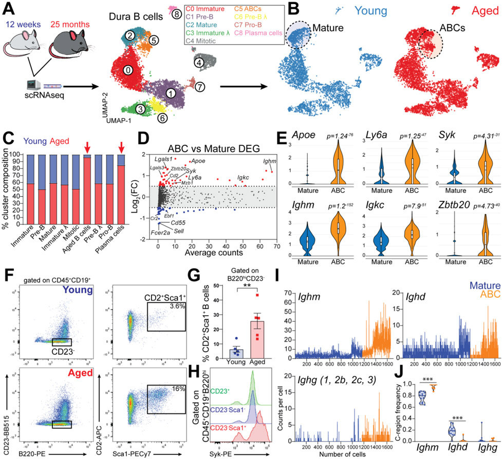
Fig. 7. Age-associated B cells disseminate throughout the dura of aged mice.
(A) Schematic depiction of the experimental design of scRNAseq comparing young and aged mice. UMAP of 9,352 B cells aggregated from seven 12-week-old and seven 25-month-old C57BL6 female mice (data generated from two independent experiments). (B) Distribution of B cells from young and aged mice. (C) Contribution of young versus aged mice to each cluster. (D) Differential gene expression analysis of ABCs versus mature B cells. (E) Violin plots showing top up-regulated genes in ABCs compared to mature B cells. (F) Dura ABCs gated as the B220hiCD23−CD2+Sca1+ cells. (G) ABC population is significantly increased in the dura of aged mice compared to young mice (mean ± SEM; n=5 mice; unpaired Student’s t test **P<0.01; data generated from two independent experiments). (H) Flow cytometry histogram showing increased levels of Syk protein in dura ABCs (representative of three 18-month-old female mice; data generated from a single experiment). (I) Ig heavy chains transcript counts per cell in ABCs and mature B cells. (J) Frequency of heavy chains usage determined by BCRseq in ABCs and mature B cells (violin plot; n=11-14 mice per group; two-ways ANOVA and Bonferroni post-hoc test ***P<0.001).

