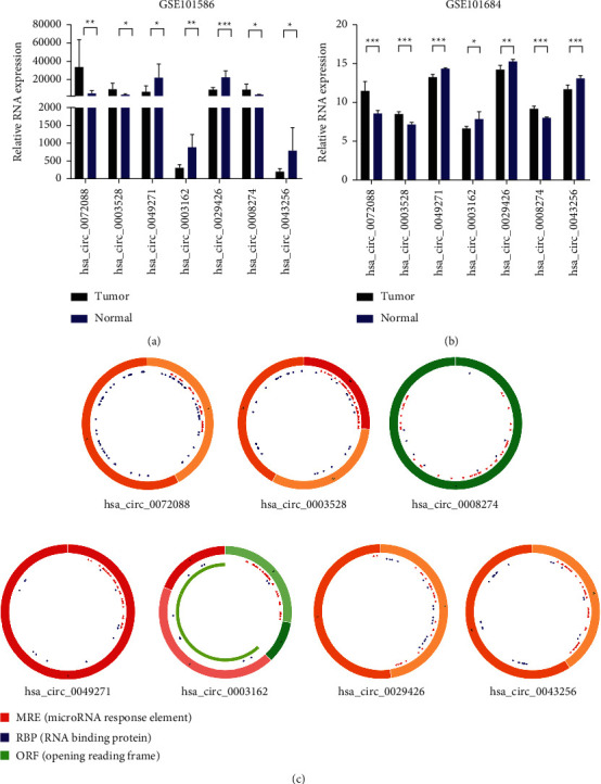
Figure 3.

Characterization of DEcircRNAs. (a, b) The expression levels of DEcircRNAs in human LUAD tissues and normal lung samples were analyzed based on GSE101586 and GSE101684 datasets. The differences were compared by paired t-test. Mean ± SEM, n = 3, ∗P < 0.05; ∗∗P < 0.01; ∗∗∗P < 0.001. (c) Structural patterns of the 7 circRNAs: hsa_circ_0072088, hsa_circ_0003528, hsa_circ_0008274, hsa_circ_0049271, hsa_circ_0003162, hsa_circ_0029426, and hsa_circ_0043256. The green part represents the open reading frame (ORF) of circRNAs. The blue part is the place where circRNA binds to the proteins. The red small triangle represents the binding position of the circRNA to the miRNA.
