
Figure 1.
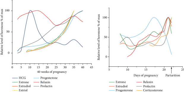
Schematic illustration of the changes in the levels of serum hCG [3], estrone [4], estradiol [4], estriol [4], progesterone [5], relaxin [6], and prolactin (PRL) [5, 7] during gestational weeks 6-40 in humans according to literature as indicated (the numbers in the x-axis indicate the gestational week). Blood samples were obtained through venipuncture of pregnant human females, and the diagnosis of pregnancy was confirmed by urine hCG determination and/or ultrasonography. HCG was determined via the double-antibody beta radioimmunoassay. Estrogens (estrone, estradiol, and estriol) were determined with an antibody against estrone-17-oxime coupled with bovine serum albumin. Progesterone was measured via the Bayer ADVIA Centaur assay. Relaxin was measured via radioimmunoassay with iodine 125-labeled polytyrosyl-relaxin and rabbit anti-porcine relaxin serum R6. PRL was determined via radioimmunoassay with homologous double antibodies. (b) Summary of the changes in the levels of estrone [8], estradiol [8], progesterone [9], relaxin [10], PRL [11], and corticosterone [12] in rat from gestational days 8-22 according to existing reports as indicated (the x-axis indicates the gestational days). The samples were derived from the peripheral blood of primiparous Sprague-Dawley rats (estrone, estradiol, progesterone, relaxin, and PRL) or the arterial blood of Wistar rats (corticosterone). Estrone and estradiol were determined via radioimmunoassay with estradiol-17β antiserum. Progesterone was quantified by protein-binding displacement. Relaxin was measured via the interpubic ligament bioassay. PRL was determined via radioimmunoassay. Corticosterone was measured using the fluorometric method.