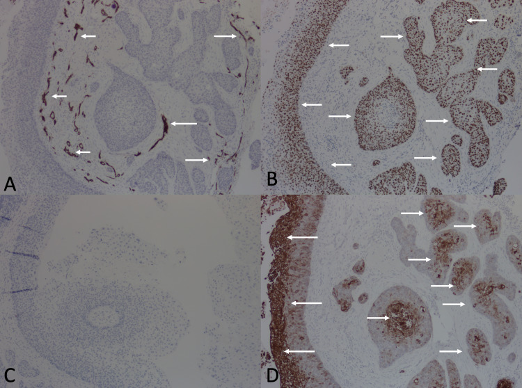
Figure 2. Immunohistochemistry of 1.5 x 1.2-centimeter biopsy of the scalp.
(A) Arrows point to areas of CD34 expression, which is limited to blood vessels (positive internal control). The tumor is otherwise negative for CD34 expression. (B) Arrows point to areas expressing p53. (C) No CK 7 expression is detected. (D) Arrows point to areas expressing CK 17.
Abbreviations: CD, cluster of differentiation; CK, cytokeratin

